
When a pet isn’t feeling well, it can be difficult to know what’s going on inside their body. However, animals cannot tell about their symptoms, unlike humans, and that is why veterinarians use the tools of the next generation to comprehend their state. Ultrasound is one of the most effective and widely used tools in contemporary veterinary care.
Pet ultrasound is an imaging method in pets that enables veterinarians to view internal organs and tissues in real-time. It is a major component in the diagnosis of diseases, checking on the health conditions, and prescriptions of treatment- without necessarily having to conduct surgery.
How Ultrasound Works
Ultrasound technology involves the use of sound waves of high frequency to produce images of the internal part of the body. A small handheld device, known as a transducer, sends these sound waves into your pet’s body. When the waves collide with internal structures such as organs or fluids they are reflected back in form of echoes.
The echoes are then translated to visual pictures on a screen. The process occurs immediately and this means that the veterinarian can monitor the movement as it takes place. To give an example, they may observe the heartbeat, observe blood circulation or monitor how organs are working in a real time.
Ultrasound does not involve the use of radiation as opposed to X-rays. It is a safer choice considering that it is needed repeatedly with time.
Why Veterinarians Use Ultrasound
Ultrasound can be frequently prescribed when a pet exhibits symptoms that cannot be detected with a basic physical examination. It gives a closer examination of the body and assists in finding out its issues that can not be seen physically.
Ultrasound is the method that veterinarians employ to research a wide variety of issues. These can involve nausea without any known cause, loss of weight, abdominal pain, abdominal swelling or behavioral change. It is also widely applied when the blood tests or any other types of diagnostic procedures indicate that there is a problem to be evaluated.
Since ultrasound provides a close-up view of soft tissues, it is specifically useful in analyzing organs and identifying abnormalities like tumors, cysts, or infections.
Which Areas of the Body of a Pet Can Be Inspected?
Ultrasound is particularly useful to see soft tissues and internal organs. It is normally employed to test the liver, kidneys, bladder, spleen, and intestines. These organs cannot be evaluated well using physical examination alone and ultrasound can be useful.
Another area that ultrasound is extensively used is in the heart. An echocardiogram is a special form of ultrasound that enables veterinarians to assess the structure and functioning of the heart. They are able to see the movement of the chambers of the heart, the opening and closing of valves and the flow of blood.
The presence of fluid in the abdomen or chest can also be detected by ultrasound and it can be an indication of underlying health issues that need to be addressed.
Ultrasound and Pregnancy in Pets
Pregnancy monitoring is one of the most popular applications of ultrasound in animals. It is applied by veterinarians to ascertain pregnancy and monitor the progress of unborn puppies, kittens or other animals.
Ultrasound gives them the opportunity to perceive the heartbeats, approximate the number of offspring and make sure that everything is moving in the right direction. It is painless and less invasive to check the mother and her babies during the pregnancy.
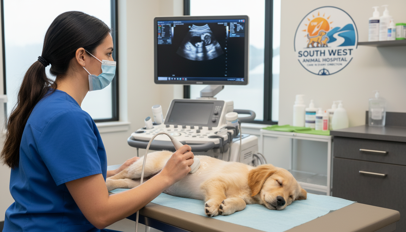
What to Expect During the Procedure
A pet ultrasound is typically not painful and is usually simple. The process is normally conducted in a serene and calm atmosphere to enable your pet to be calm.
Your pet will be requested to lie on a table, it can be on his or her back or side, depending on the part that is under examination. A small area of fur is in most cases shaved to enable closer contact between the skin and the transducer. This assists in creating better images.
The skin is applied with a special gel. Such gel can have a bit of a cool feel, but it is non-invasive and, again, it assists the sound waves to work better. The veterinarian then rubs the transducer over the area to get images.
Most pets are not bothered by the procedure and do not move. No needles or incisions are required and the procedure is painless unless a guided procedure is being done.
Do Pets Have to be Sedated to Ultrasound?
Sedation is not necessary in most cases. Because the procedure is comfortable and not invasive, most pets are calm enough to be without medication.
In specific cases, however, when a pet is unusually nervous, agitated or suffering, mild sedation can be prescribed. This makes sure that the images are not affected and the procedure can be done safely without stressing the animal.
Conditions That Ultrasound Can Help Detect
Ultrasound is an immense diagnostic device capable of detecting a broad range of health problems. It is able to demonstrate tumors, cysts, enlargement of organs, and internal bleeding. It also comes in handy to identify obstructions in the digestive or urinary system.
Ultrasound imaging can also be used to identify infections and inflammation in organs. As an illustration, it may be used to diagnose liver, kidney, or pancreatic conditions.
Moreover, ultrasound may be employed to assist in some medical operations. Veterinarians can use it to help in biopsies or fluid collection, enabling them to insert needles at the correct location and safely.
How Ultrasound Differs from Other Imaging Methods
Ultrasound is very effective, although it is usually employed together with other imaging. Both approaches have their merits.
X-rays are more suitable when it comes to analyzing bones or when it is necessary to determine the presence of a fracture or some other issue with the lungs. Ultrasound in contrast is very good in visualization of soft tissues and fluid structures.
Veterinarians in most instances use both techniques in order to have comprehensive information about the health of a pet. Such a holistic practice will result in improved diagnoses and treatment.
How Long Does the Procedure Take?
The duration of an ultrasound session may be different based on the areas that have to be investigated. A majority of the procedures last between 20 and 45 minutes.
In case a more in-depth assessment is necessary, it might require a bit more time. In other instances the veterinarian can examine the pictures further or even discuss with a specialist and then give a final diagnosis.
Is Ultrasound Safe for Pets?
It is believed to be one of the safest diagnostic tests that can be performed on animals. Since it does not use radiation, it can be done several times.
This test is painless, non-invasive, and tolerated well by most animals. Thus, it is highly recommended for diagnosis purposes.
Closing Thought
Ultrasound for pets has become an essential part of modern veterinary care, offering a safe, painless, and highly effective way to understand what’s happening inside your pet’s body. It allows veterinarians to detect problems early, monitor health conditions, and make informed treatment decisions without the need for surgery. While it may seem like a simple procedure, its impact on your pet’s health and well-being is significant. By using advanced tools like ultrasound, pet owners can ensure their companions receive timely, accurate, and compassionate care.
At South West Animal Hospital we are committed to providing advanced diagnostic services and expert care to help your pets live healthier and happier lives.